Rivercyte - fostering the future of cell analysis through deformability cytometry.
The future of cell analysis.
At Rivercyte, we produce technology for the analysis of physical phenotypes of cells, including their deformability - information largely missing in current biological and medical research. We envision that in the future, insights about the physical phenotype of cells will be informing decisions in clinical practice.
We democratize deformability cytometry. We make it easy-to-use and affordable. This way, we hope to make the technology widely available.
Our Technology
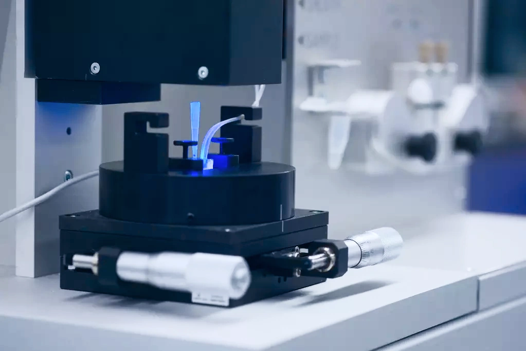
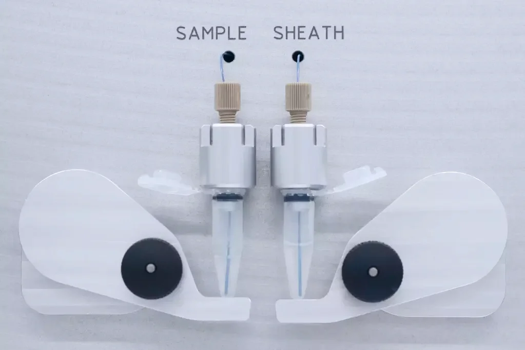
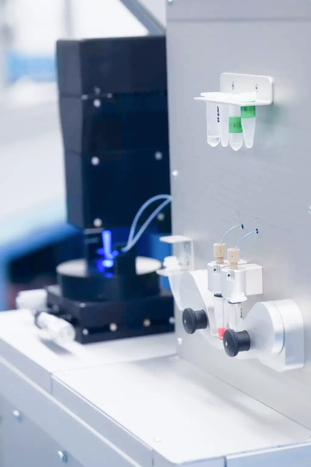
We produce devices and software for the analysis of physical properties of cells, including their stiffness, size, shape and morphological features.
Why do the mechanical properties of cells matter?
Cell mechanics and other physical properties of cells reflect physiological and pathological changes in cell function. They are promising biomarkers for disease diagnosis and prognosis. For example, we recently found dramatic changes of blood cell mechanics during COVID-19.
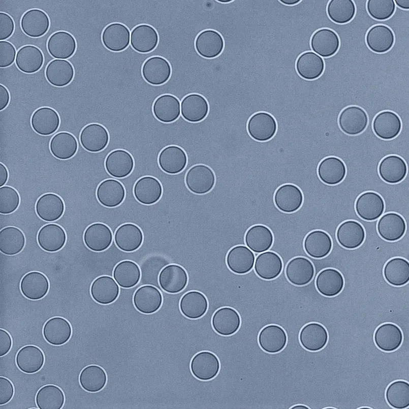
An HL-60 cell deforming under shear stress in a microfluidic channel. Video by MPL, licensed CC0.
What is physical phenotyping of cells?
Physical phenotyping involves the characterization of single living cells by their physical and morphological features such as size, shape and stiffness. An efficient technique for doing this is deformability cytometry. As a label-free method based purely on brightfield images of cells, deformability cytometry adds a new functional and unbiased dimension to flow cytometry. Combined with machine learning, deformability cytometry is a discovery machine for characterizing cell populations and functional states that are invisible to marker-based techniques.
What is deformability cytometry?
In deformability cytometry, thousands of single cells flow quickly through a narrow microfluidic channel, where the cells deform due to the forces applied. An image of each cell is taken and analyzed. Cell deformability and other parameters describing the cell's physical phenotype are extracted from the image. Thanks to high-speed video microscopy, our analysis rates of 1000 cells per second approach those of conventional fluorescence-based flow cytometers. Hundreds of thousands of cells are measured within minutes.
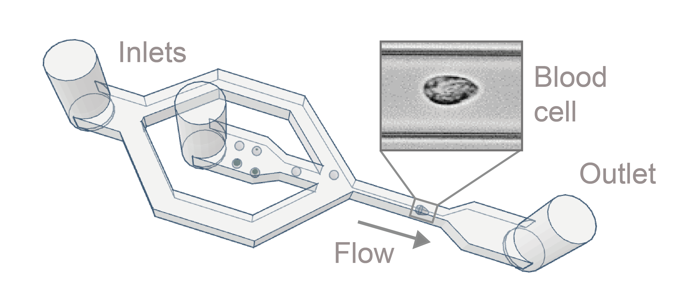
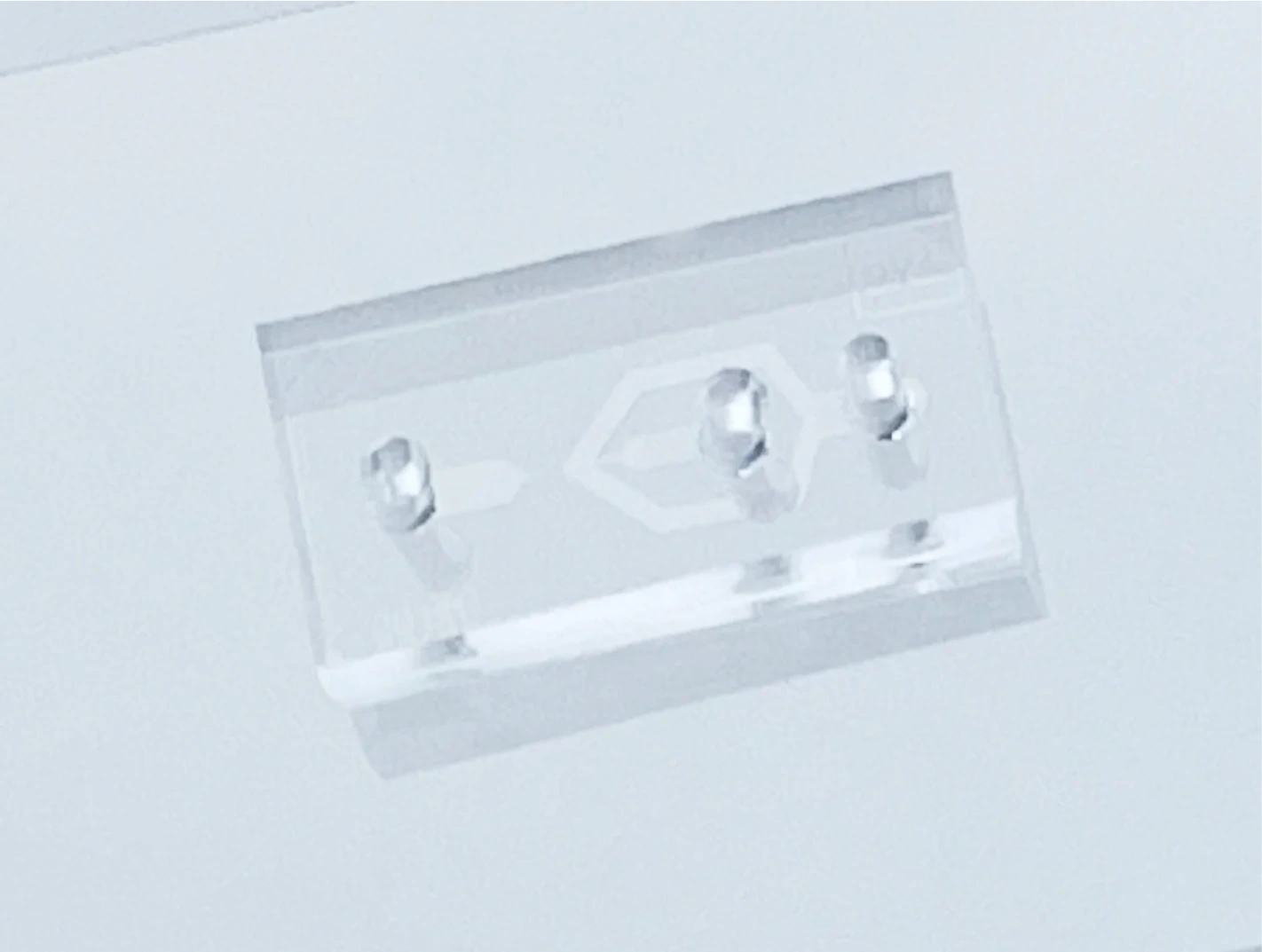
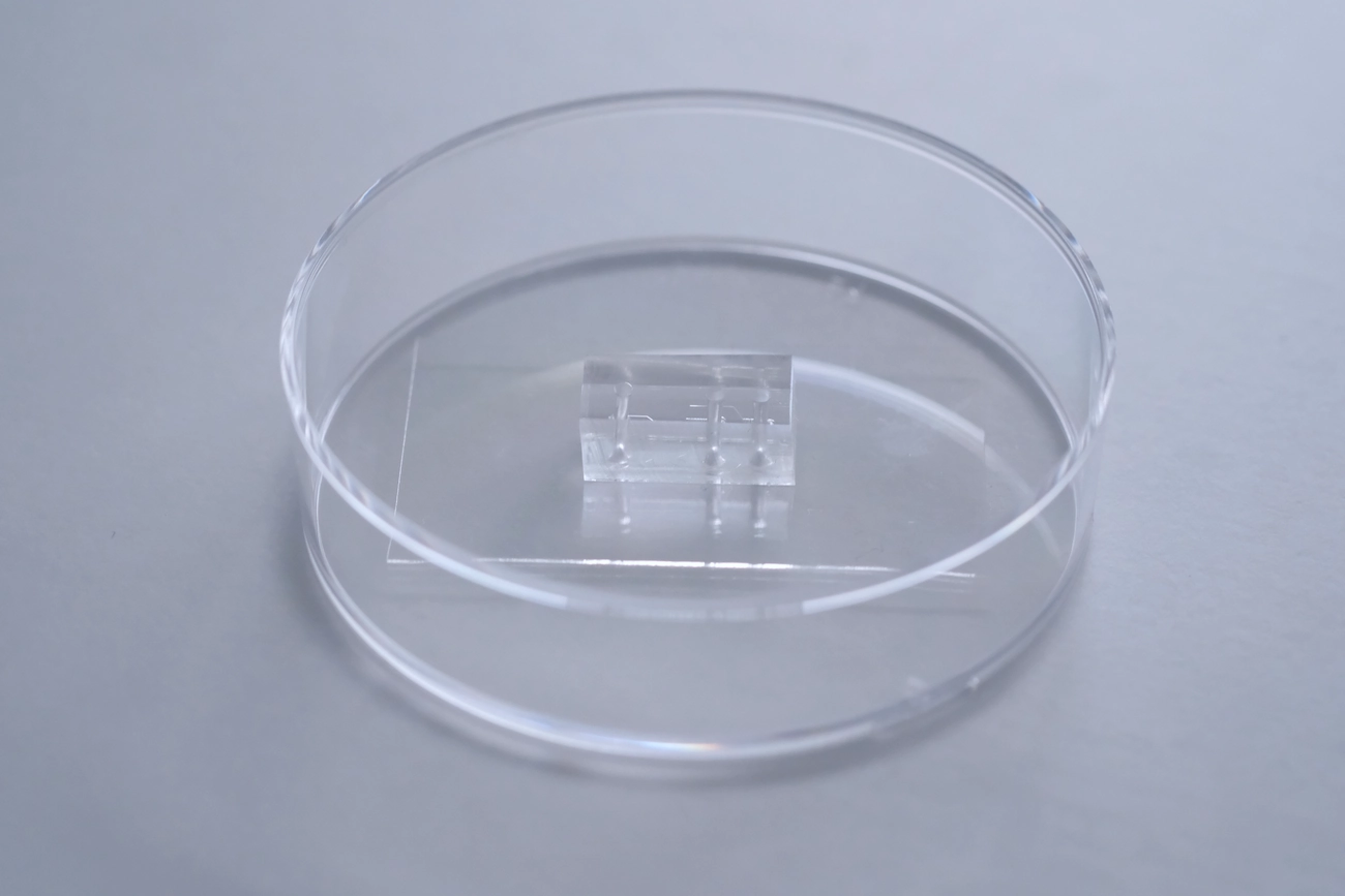
Scheme of the microfluidic chip used for deformability cytometry. Image by MPL, licensed CC0.
Which samples can be analyzed?
Any cell suspension can be measured with deformability cytometry - blood and other liquid biopsies, cells from solid tissues, and cultured cells. All of this is possible without time- and material-consuming sample preparation such as staining. As such, deformability cytometry supports diverse applications in biology, biotechnology and medicine.
Key publications from our team
- Otto et al., Real-time deformability cytometry: on-the-fly cell mechanical phenotyping. Nature Methods (2015)
- Rosendahl et al., Real-time fluorescence and deformability cytometry. Nature Methods (2018)
- Toepfner et al., Detection of human disease conditions by single-cell morpho-rheological phenotyping of blood. eLife (2018)
- Nawaz, Urbanska and Herbig et al., Intelligent image-based deformation-assisted cell sorting with molecular specificity. Nature Methods (2020)
- Urbanska et al., A comparison of microfluidic methods for high-throughput cell deformability measurements. Nature Methods (2020)
- Kubánková et al., Physical phenotype of blood cells is altered in COVID-19. Biophysical Journal (2021)
- Soteriou and Kubánková et al., Rapid single-cell physical phenotyping of mechanically dissociated tissue biopsies. Nature Biomedical Engineering (2023)
News

Rivercyte presents at MedtechSUMMIT 2024
2024-05-15Our founders Shada Hofemeier Abuhattum and Martin Kräter presented the Naiad and our data analysis solutions at MedtechSUMMIT 2024 in Regensburg. The event, centered on "Digitale Diagnostik - Die transformative Kraft von KI in der Diagnostik?", highlighted AI's transformative impact on digital diagnostics, discussing challenges and innovative solutions.

Rivercyte showcases the Naiad at the “Mechanics of Life” symposium at EMBL Heidelberg
2024-04-18Rivercyte showcased their Naiad device at the "Mechanics of Life" symposium at EMBL Heidelberg, demonstrating their deformability cytometry technology. The symposium, focused on the role of mechanical forces from molecules to organs in development and disease, was attended by an interdisciplinary audience. Rivercyte cofounders Martin Kräter and Markéta Kubánková presented projects on deformability cytometry, including a method for diagnosing colon cancer biopsies using AI and TissueGrinder.

Rivercyte partners with leading institutions on innovative neonatal diagnostics project
2024-03-01Rivercyte is collaborating with University Hospital Erlangen and the Max Planck Institute for the Science of Light on the SoPhyA project, supported by the Bavarian State Ministry. Over three years, the project aims to enhance neonatal diagnostics using deformability cytometry to study blood cell mechanics in pediatric patients. This innovation seeks to improve early health detection and outcomes in neonatal care, supported by BayVFP funding of €319,000.
Products
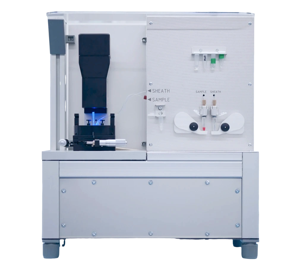
Deformability Cytometry Device and Software
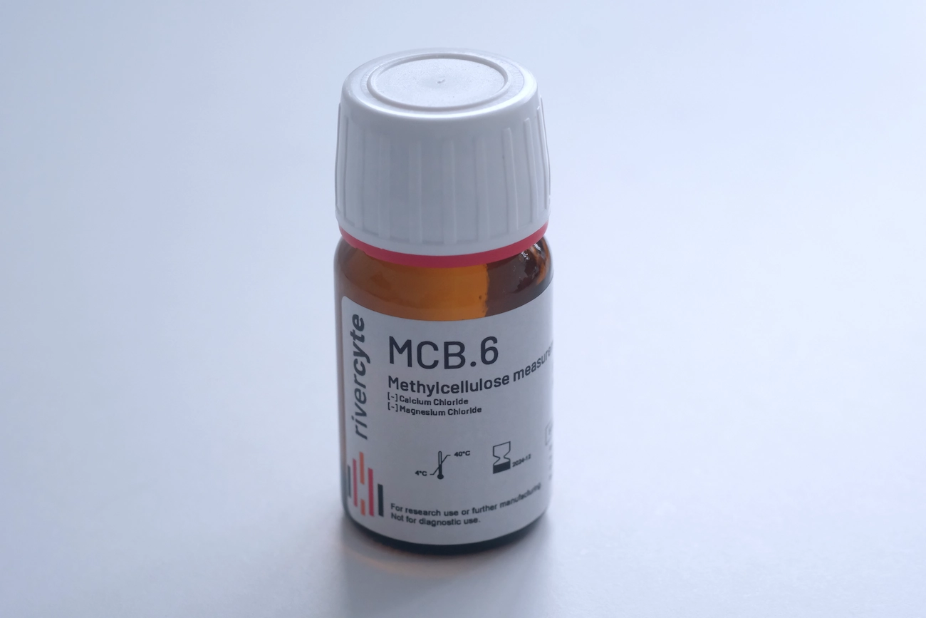
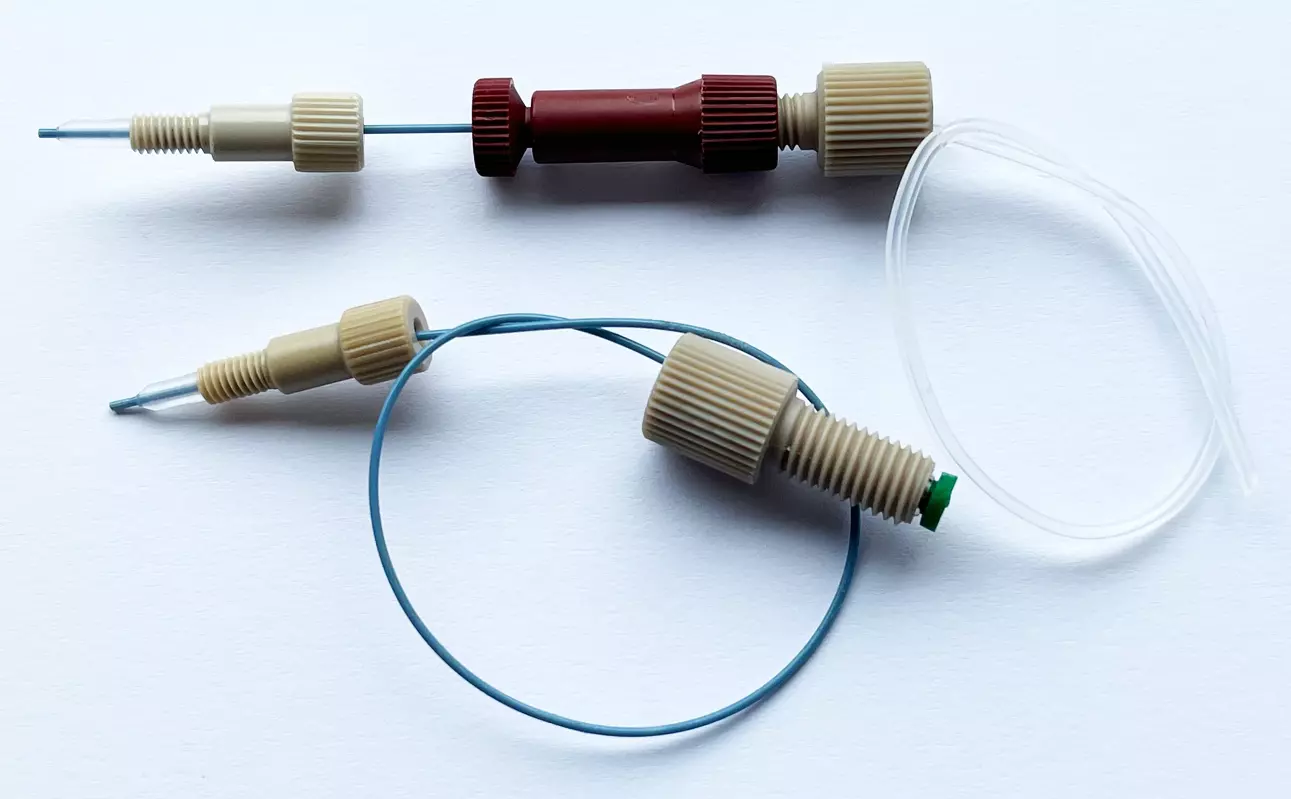
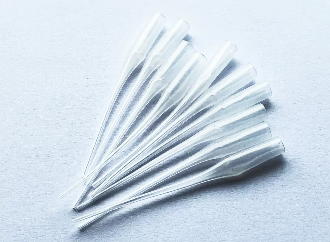
Consumables for Deformability Cytometry
